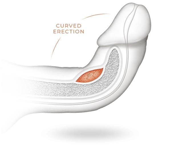
Peyronie’s disease is a wound-healing disorder in which fibrous plaque forms in the tunica albuginea of the penis, causing curvature, shortening, an hourglass deformity, pain, and often erectile dysfunction. Treatment depends on whether the disease is still active, how severe the bend is, and whether erections are still adequate.
Also referred to as: penile curvature, induratio penis plastica, bent penis correction, penil eğrilik, penile plaque surgery.
What is happening inside the penis
The erectile bodies are wrapped in a tough elastic sheath called the tunica albuginea. In Peyronie’s disease, an area of that sheath is replaced by inelastic scar — a plaque. When the penis fills with blood, the healthy tunica stretches and the plaque does not, so the shaft bends toward the plaque, narrows at the level of the plaque, or both. The plaque is not a tumour and it is not cancer.
It usually begins after an unnoticed injury: a bend during intercourse, a straddle injury, or repeated micro-trauma. Some men have a genetic predisposition to abnormal wound healing, which is why Peyronie’s runs alongside Dupuytren’s contracture of the hand and plantar fascia disease in the same families. Diabetes, smoking and low testosterone increase the risk. Around one man in twenty is affected, and the true figure is almost certainly higher because so many never present.
The distinction that governs everything else is the phase of the disease. In the active phase — typically the first six to eighteen months — the plaque is still forming, the curve is still changing, and erections are often painful. In the stable phase the plaque has matured, pain has resolved, and the curvature has not changed for at least three months. Surgery belongs to the stable phase; operating on an active plaque invites recurrence.
How the deformity is assessed
Accurate correction depends on accurate measurement. A photograph of a partial erection is not enough to plan surgery on.
- History: date of onset, whether the curve is still changing, presence of pain, and the effect on penetration
- Validated questionnaires — IIEF-5 for erectile function and the PDQ for bother and symptom severity
- Examination of the flaccid penis to map plaque position, size and any calcification
- An induced erection using an intracavernosal injection, so the curvature can be measured with a goniometer in degrees rather than estimated
- Doppler ultrasound during the same injection to assess arterial inflow, venous leak and plaque calcification
- Stretched penile length, documented before any decision is made — because every straightening operation trades a little length for a straight shaft
Non-surgical treatment in the active phase
If the disease is still evolving, the aim is to control pain, limit the final deformity and buy time until the plaque matures. Nothing reliably dissolves an established plaque, and any clinic promising otherwise is overselling.
Penile traction therapy
The only conservative measure with consistent evidence for both curvature reduction and length preservation. It requires genuine commitment — several hours a day over three to six months — and we set that expectation openly rather than handing over a device and hoping.
Intralesional collagenase
Collagenase clostridium histolyticum injected directly into the plaque can reduce curvature by roughly 30–35% in selected patients with a bend between 30° and 90° and no calcification. Availability varies by country and it is not a substitute for surgery in severe deformity.
Intralesional verapamil
A lower-cost injectable option with more modest and less consistent evidence, sometimes combined with traction. Reasonable where collagenase is unavailable and the patient wants to try a conservative course first.
Shockwave therapy for pain
Low-intensity extracorporeal shockwave treatment reduces the pain of the active phase in many men. It does not straighten the penis, and we say so before anyone pays for a course of it.
Choosing the right operation
Three surgical strategies exist, and the choice is dictated by the degree of curvature, the quality of erections, and penile length. Getting that choice right matters more than the technical polish of any individual technique.
| Technique | Indicated when | What it involves | Trade-off |
|---|---|---|---|
| Plication (Nesbit, Yachia, 16-dot) | Curvature under about 60°, good rigidity, no hourglass narrowing, adequate length | Shortening the longer, healthy side of the tunica with sutures so the shaft straightens. The plaque itself is left alone. | Loses roughly 1–2 cm of length. Simplest operation, lowest risk to erectile function, sutures may be palpable. |
| Plaque incision or partial excision with grafting | Curvature over 60°, hourglass or hinge deformity, but erections still good without medication | The plaque is cut to release the tethering, and the resulting tunical defect is patched with a graft such as bovine or processed pericardium. | Preserves length. Carries a 10–25% risk of worsened erectile function, so it is reserved for men with genuinely strong erections. |
| Penile implant with straightening | Curvature of any degree combined with erectile dysfunction that no longer responds to medication | An inflatable prosthesis is placed, and the shaft is straightened over it by manual modelling, plaque incision or grafting as required. | Solves curvature and erectile dysfunction in one operation. Irreversible, as with any implant. |
A residual curve of up to 20° is functionally irrelevant and is accepted in all three techniques rather than chased with additional risk.
Surgical atlas
The two operations, step by step
Both straightening operations are drawn below from the surgeon’s own viewpoint: the lateral view for what is being measured, the dorsal or ventral operative field for what is being done. Step through them to see where the plaque sits, what is cut, and what is left behind.
Plaque incision with grafting
Release the scar, patch the hole, keep the length.
Indicated when Curvature over about 60 degrees, an hourglass or hinge deformity, and erections still rigid without medication.
-
Step 1 of 6 Lateral view — erect
Artificial erection and goniometry
Saline is infused into the corpora on the table so the erection behaves exactly as it does at home, and the bend is then measured with a goniometer rather than estimated by eye. The hourglass narrowing at the level of the plaque is recorded in the same breath, because narrowing is what rules plication out.
A curve over 60 degrees with an hourglass waist and strong erections is the classic indication for grafting rather than shortening the opposite side.
- Measured curve
- 75°
- Deformity
- Hourglass
- Erections
- Rigid, no tablets
-
Step 2 of 6 Dorsal exposure
Circumcising incision and degloving
A circumcising incision below the corona lets the skin and dartos be rolled back to the base as a single sleeve, which exposes Buck’s fascia over the whole length of the shaft. The sleeve is held out on stay sutures. Nothing is excised, and the foreskin is preserved in men who are not already circumcised.
Working through a full degloving rather than a local incision is what makes it possible to see the whole plaque, and to check the correction at the end without a second approach.
- Access
- Subcoronal, circumferential
- Preserved
- Foreskin and skin sleeve
-
Step 3 of 6 Dorsal exposure
Elevating the neurovascular bundle
Buck’s fascia is opened lateral to the deep dorsal vein and the sheet carrying the dorsal nerves and arteries is lifted off the tunica as one layer, then held aside. Only now is the plaque bare and safe to cut.
This is the step that protects sensation. The dorsal nerves lie directly on the plaque, and a relaxing incision made before they are elevated is how numbness happens.
- Elevated
- Nerves, arteries, dorsal vein
- Plane
- Sub-Buck’s, off the tunica
-
Step 4 of 6 Dorsal exposure
Relaxing incision through the plaque
A transverse incision is carried through the plaque at the point of maximum tethering, with a short relaxing limb at each end — the H incision. The instant the scar is divided the short side of the shaft lengthens and the penis comes straight, leaving a defect in the tunica that has to be filled.
The defect is deliberately not closed directly. Pulling it shut would simply re-create the tether and hand back the curve.
- Incision
- Transverse H
- Defect
- Approx. 30 x 20 mm
- Length
- Restored on the short side
-
Step 5 of 6 Dorsal exposure
Grafting the tunical defect
The defect is measured and a graft — processed bovine pericardium in most of our cases — is cut about a fifth larger than the gap and sewn in with a continuous fine monofilament. The oversizing is not cosmetic: a graft cut to the exact size of the defect contracts as it heals, and the curve comes back with it.
Pericardium is used because it is acellular, takes a suture without tearing, and has no donor site. A vein graft harvested from the leg is the alternative and costs an extra incision.
- Graft
- Processed pericardium
- Oversized by
- Approx. 20%
- Closure
- Continuous 4-0 monofilament
-
Step 6 of 6 Lateral view — erect
Straightness check and closure
A second artificial erection confirms the correction before a single layer is closed. The bundle is then laid back over the graft, Buck’s fascia is repaired, and the skin sleeve is drawn forward again. A residual bend of under 20 degrees is accepted rather than chased with more dissection.
Length is preserved, and often a few millimetres are recovered from the side the plaque had been holding short. Gentle stretching starts at about four weeks to keep the graft supple.
- Residual curve
- 5°
- Length
- Preserved
- Plaque
- Divided, graft in place
Nesbit plication
Shorten the long side until the shaft is straight.
Indicated when Curvature under about 60 degrees, no narrowing, good rigidity, and enough length to spend on the correction.
-
Step 1 of 5 Lateral view — erect
Artificial erection and goniometry
The same measured start: saline induces the erection, the goniometer gives a number, and the shaft is checked for narrowing. A curve of about 40 degrees with no waist, good rigidity and length to spare is what plication is for.
Stretched length is written down before anything else happens, because this operation spends length to buy straightness and the patient has to agree to that trade in advance.
- Measured curve
- 40°
- Deformity
- Simple dorsal bend
- Erections
- Rigid, no tablets
-
Step 2 of 5 Ventral exposure
Exposing the convex side
The correction is made on the outside of the bend, not on the plaque. For a dorsal curve that means working ventrally, so after degloving the corpus spongiosum carrying the urethra is mobilised off the tunica and slung clear of the segment that is to be shortened.
The urethra has to be lifted rather than worked around. A suture placed through the spongiosum is the complication nobody wants to explain afterwards.
- Working surface
- Ventral tunica
- Mobilised
- Corpus spongiosum
- Plaque
- Not touched
-
Step 3 of 5 Ventral exposure
Paired elliptical excisions
Transverse ellipses of tunica are marked either side of the urethra at the point of maximum convexity, sized at roughly a millimetre of width for every degree of bend, and excised full thickness. Pairing them keeps the shortening symmetrical.
Taking the whole correction from one side trades an upward curve for a sideways one. The Yachia variant makes a longitudinal incision closed transversely instead, and the 16-dot technique gathers the tunica with sutures and excises nothing at all.
- Excisions
- Paired, transverse
- Width
- Approx. 1 mm per degree
- Depth
- Full-thickness tunica
-
Step 4 of 5 Ventral exposure
Closure with non-absorbable sutures
Each defect is closed transversely with inverted non-absorbable sutures, so the knots sit inside the tunica rather than under the skin where they can be felt. The urethra stays slung clear until both repairs are tied, then drops back into the midline over them. The convex side is now shorter than it was, and the shaft straightens without the plaque having been touched.
Palpable knots are the commonest complaint after plication, and inverting them is the whole answer. Absorbable sutures are not used here: the repair has to hold for good.
- Suture
- 2-0 non-absorbable
- Knots
- Inverted, inside the tunica
- Plaque
- Left in place
-
Step 5 of 5 Lateral view — erect
Straightness check
A repeat artificial erection confirms a straight shaft before closing. The plaque is still there, because plication never removes it, and the shaft is roughly one to two centimetres shorter than it was. That is the price of the simplest and least risky of the three operations.
Erectile function is essentially untouched by plication, which is why it stays the first choice whenever the curve is moderate and there is length to spend.
- Residual curve
- 4°
- Length
- Approx. 1.5 cm shorter
- Plaque
- Still present
Drawn from operative landmarks rather than traced from a photograph, and schematic rather than to scale. The plates show the principle of each step; the size of the plaque, the shape of the defect and the amount of tunica taken differ in every man. Send your measurements for a written opinion →
Recovery after straightening surgery
- Day 0–1 One night in hospital with a light compressive dressing. Pain is moderate and controlled with oral analgesia. The catheter, where used, is removed the following morning.
- Days 2–7 Swelling and bruising of the shaft are expected and can be dramatic in appearance without being significant. Wound review before you fly home.
- Weeks 2–4 Bruising resolves. Desk work resumes within seven to ten days. Nocturnal erections may be uncomfortable initially; this settles.
- Weeks 4–8 Where a graft has been used, we start a gentle stretching or vacuum protocol at around four weeks to keep the graft supple and preserve length.
- From week 6–8 Intercourse resumes after review. Final cosmetic result is judged at three months, once all swelling has settled.
Questions we are asked about peyronie’s disease
Will Peyronie’s disease go away on its own?
Rarely. Spontaneous improvement occurs in roughly one man in eight; in most the curvature stabilises and persists, and in a minority it worsens. Pain, in contrast, resolves by itself in the great majority of cases within twelve to eighteen months.
Will surgery make my penis shorter?
Plication deliberately shortens the longer side, so it costs roughly one to two centimetres. Grafting is designed to preserve length and sometimes restores a little of what the plaque took. We measure and document stretched length at consultation so the conversation is based on numbers rather than impressions.
Can the curvature come back after surgery?
Recurrence is uncommon when surgery is performed in the stable phase, which is precisely why we insist on stability first. Operating on an active plaque significantly increases the chance of the deformity re-forming.
Can Peyronie’s disease be treated without surgery?
In the active phase, yes — traction therapy, intralesional injections and pain management can limit the final deformity. Once the plaque is mature and the bend prevents intercourse, surgery is the only treatment that reliably straightens the penis.
I have both curvature and erectile dysfunction. What then?
That combination is common and is usually best treated with an inflatable implant plus straightening in a single operation. Correcting only the curve in a man who cannot achieve a rigid erection leaves him with a straight penis he still cannot use.
Does Peyronie’s disease increase the risk of cancer?
No. The plaque is scar tissue in the tunica albuginea. It has no malignant potential and does not develop into cancer.
Clinically reviewed by the Santa Urology surgical team. This page describes typical outcomes reported in the surgical literature and in our own practice; individual results vary and nothing here is a substitute for a personal consultation. See our medical disclaimer.